Welcome to the Amira-Avizo Software Use Case Gallery
Below you will find a collection of use cases of our 3D data visualization and analysis software. These use cases include scientific publications, articles, papers, posters, presentations or even videos that show how Amira-Avizo Software is used to address various scientific and industrial research topics.
Use the Domain selector to filter by main application area, and use the Search box to enter keywords related to specific topics you are interested in.
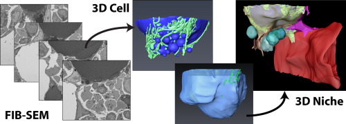
Characterization of the bone marrow adipocyte niche with three-dimensional electron microscopy
Unlike white and brown adipose tissues, the bone marrow adipocyte (BMA) exists in a microenvironment containing unique populations of hematopoietic and skeletal cells.
To study this microenvironment at the subcellular level, we performed a three-dimensional analysis of the ultrastructure of the BMA niche with focused ion beam scanning electron microscopy (FIB-SEM). This revealed that BMAs display hallmarks of metabolically active cells including polarized lipid deposits, a dense mitoch... Read more
Hero Robles, SungJae Park, Matthew S. Joens, James A.J. Fitzpatrick, Clarissa S. Craft, Erica L. Scheller
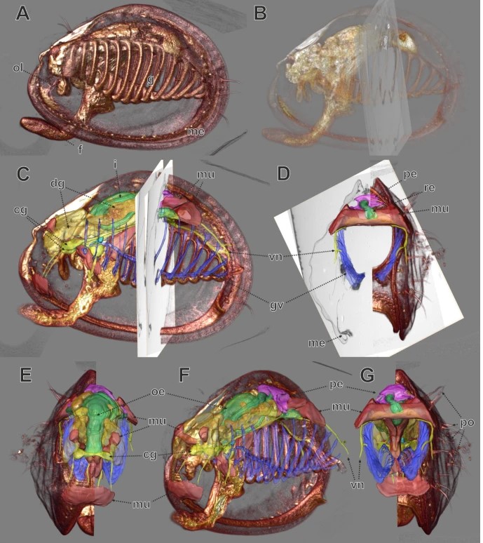
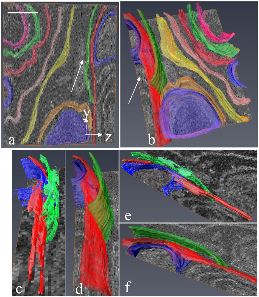
In biomedical research, a huge variety of different techniques is currently available for the structural examination of small specimens, including conventional light microscopy (LM), transmission electron microscopy (TEM), confocal laser scanning microscopy (CLSM), microscopic X-ray computed tomography (microCT), and many others. Since every imaging method is physically limited by certain parameters, a correlative use of complementary methods often yields a significant broader range of inform... Read more
Stephan Handschuh, Natalie Baeumler, Thomas Schwaha & Bernhard Ruthensteiner
As key functional units in neural circuits, different types of neuronal synapses play distinct roles in brain information processing, learning, and memory. Synaptic abnormalities are believed to underlie various neurological and psychiatric disorders. Here, by combining cryo-electron tomography and cryo-correlative light and electron microscopy, we distinguished intact excitatory and inhibitory synapses of cultured hippocampal neurons, and visualized the in situ 3D organization of ... Read more
Chang-Lu Tao, Yun-Tao Liu, Rong Sun, Bin Zhang, Lei Qi, Sakar Shivakoti, Chong-Li Tian, Peijun Zhang, Pak-Ming Lau, Z. Hong Zhou and Guo-Qiang Bi
Corpora amylacea are cell-derived structures that appear physiologically in the aged human brain. While their histological identification is straightforward, their ultrastructural composition and microenvironment at the nanoscale have remained unclear so far, as has their relevance to aging and certain disease states that involve the sequestration of toxic cellular metabolites. Here, we apply correlative serial block-face scanning electron microscopy and transmission electron tomograp... Read more
Paula P. Navarro, Christel Genoud, Daniel Castaño-Díez, Alexandra Graff-Meyer, Amanda J. Lewis, Yvonne de Gier, Matthias E. Lauer, Markus Britschgi, Bernd Bohrmann, Stephan Frank, Jürgen Hench, Gabriel Schweighauser, Annemieke J. M. Rozemuller, Wilma D. J. van de Berg, Henning Stahlberg & Sarah H. Shahmoradian
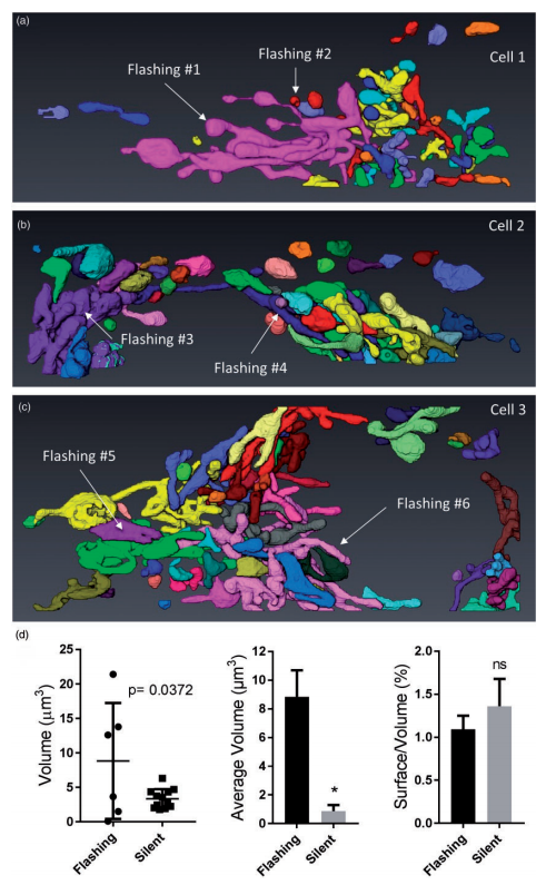
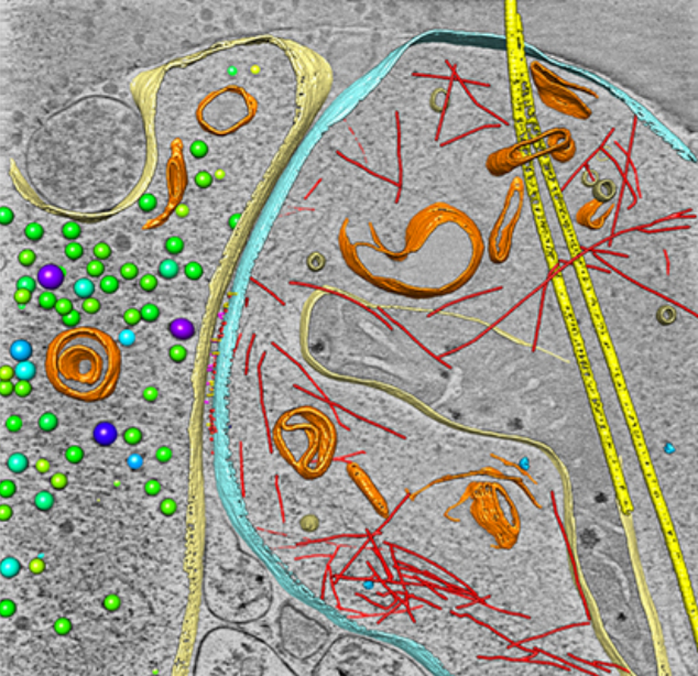
Ultrastructural Characterization of Flashing Mitochondria
Mitochondria undergo spontaneous transient elevations in matrix pH associated with drops in mitochondrial membrane potential. These mitopHlashes require a functional respiratory chain and the profusion protein optic atrophy 1, but their mechanistic basis is unclear. To gain insight on the origin of these dynamic events, we resolved the ultrastructure of flashing mitochondria by correlative light and electron microscopy. HeLa cells expressing the matrix-targeted pH probe mitoSypHer were screen... Read more
Manon Rosselin, Paula Nunes-Hasler, and Nicolas Demaurex
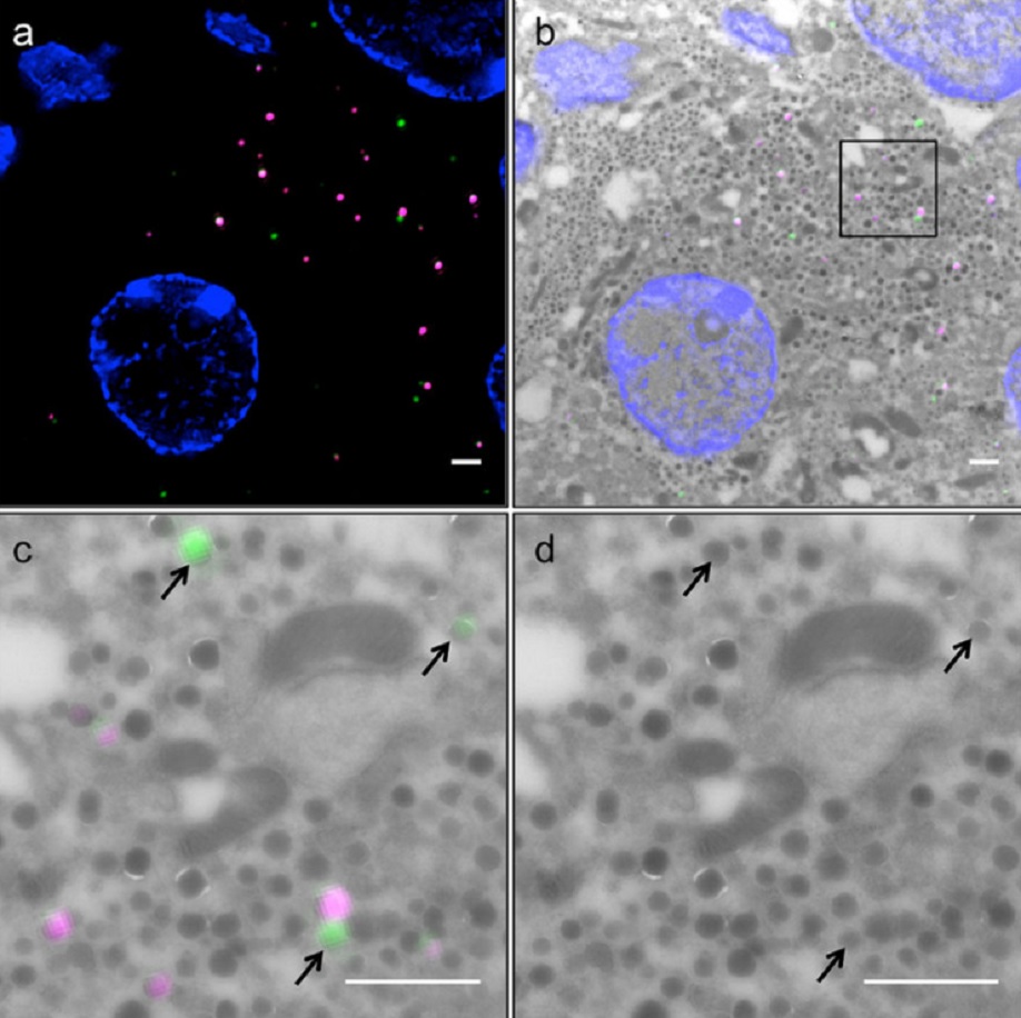
Correlative light and electron microscopy (CLEM) is a powerful approach to investigate the molecular ultrastructure of labeled cell compartments. However, quantitative CLEM studies are rare, mainly due to small sample sizes and the sensitivity of fluorescent proteins to strong fixatives and contrasting reagents for EM. Here, we show that fusion of…
Read more
Andreas Müller, Martin Neukam, Anna Ivanova, Anke Sönmez, Carla Münster, Susanne Kretschmar, Yannis Kalaidzidis, Thomas Kurth, Jean-Marc Verbavatz & Michele Solimena
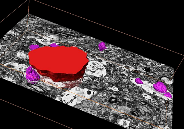
Serial block-face electron microscopy (SBEM) provides nanoscale 3D ultrastructure of embedded and stained cells and tissues in volumes of up to 107 µm3. In SBEM, electrons with 1–3 keV energies are incident on a specimen block, from which backscattered electron (BSE) images are collected with x, y resolution of 5–10 nm in the block-face plane, and successive layers are removed by an in situ ultramicrotome. Sp... Read more
Q. He, M. Hsueh, G. Zhang, D. C. Joy & R. D. Leapman
Volcanogenic Pseudo-Fossils from the ∼3.48 Ga
The ∼3.48 billion-year-old Dresser Formation, Pilbara Craton, Western Australia, is a key geological unit for the study of Earth’s earliest life and the habitats it occupied. Here, we describe a new suite of spheroidal to lenticular microstructures that morphologically resemble some previously reported Archean microfossils. Correlative microscopy shows that these objects have a size distribution, wall ultrastructure, and chemistry that are incompatible with a microfossil origin and in... Read more
Wacey David , Noffke Nora , Saunders Martin , Guagliardo Paul , and Pyle David M